Know the moment your cells change.
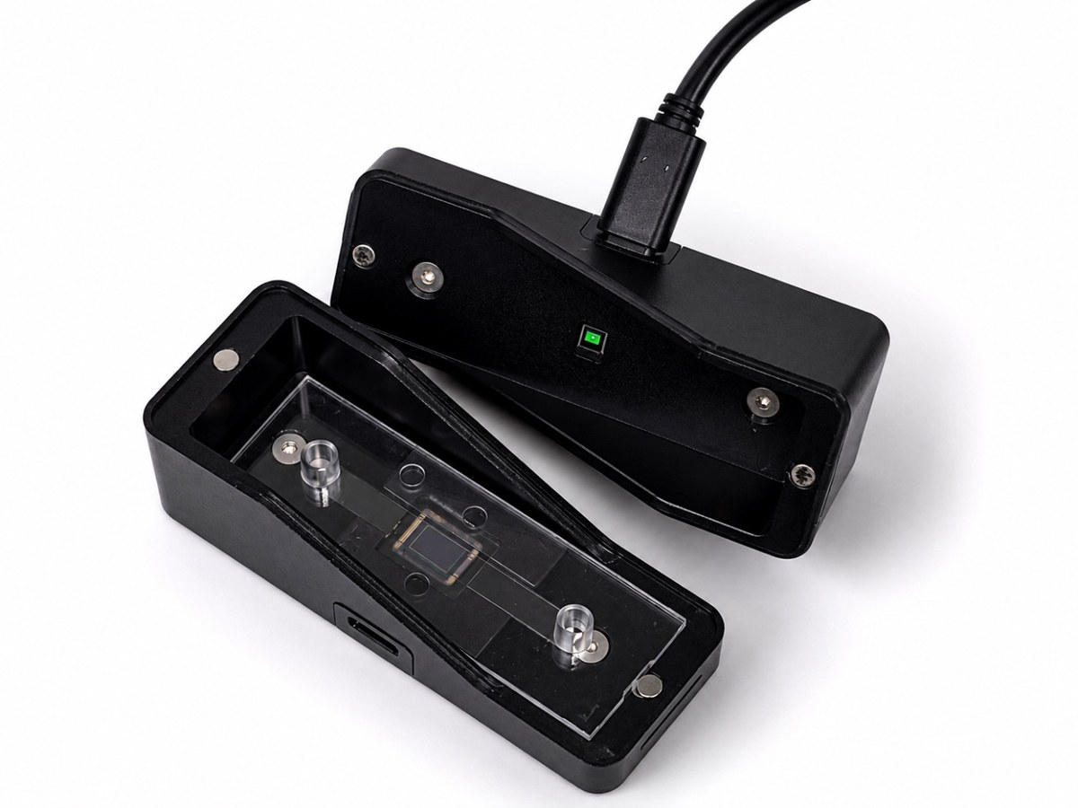
A microscope small enough to live inside the incubator, measuring the culture around the clock.
Working prototype, validated in published work.
We are building the productised instrument with a small number of research partners.
TALK TO US ABOUT YOUR SAMPLES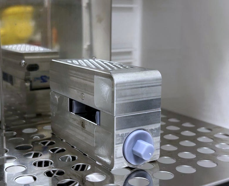
01What it is
Live-cell imaging systems can already hold a culture under control for days. The limitation XipScope addresses is not getting into the incubator — it is that one optical system has to serve every sample, so wells are visited in turn and a culture that should be watched continuously is watched in rotation.
XipScope is a distributed architecture instead. Each sample gets its own compact module, fixed on the incubator shelf beside it. With no moving parts, nothing scans and nothing waits its turn: scaling means adding modules, not building a faster stage.
The optics do not care what the sample is. The same instrument has been used on angiogenesis assays, on developmental biology and on wine fermentation. We are pointing it at organ-on-a-chip, because those assays run in plates — and a plate is exactly what a per-module architecture is for. The design is moving toward one module per well.
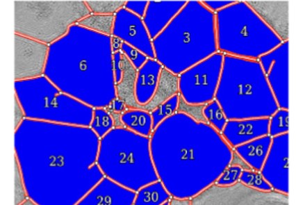
The aim is not to give you more images. It is to give you the number the experiment depends on — tube length, junction count, cell density, or whatever yours measures — and to flag the moment it changes. Once the structure is traced, those numbers come out on their own.
Every assay reads a different structure, so we build the tracing for yours. That is why we work with a few groups at a time, rather than shipping one tool that measures everything approximately.
02What it sees
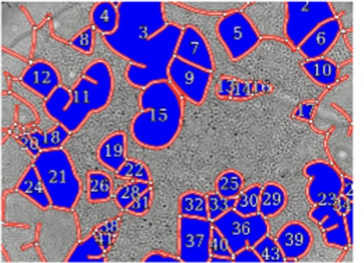
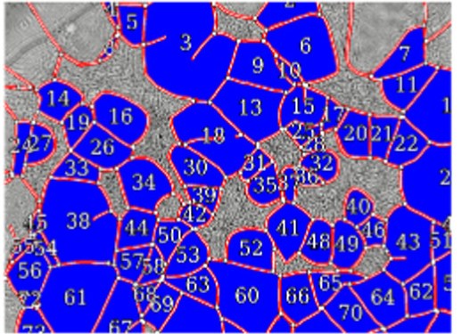
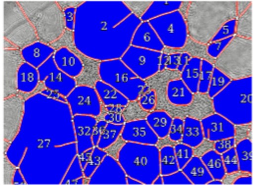
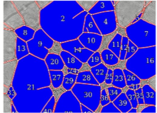
Everything below was imaged with the culture left where it was.
Against the benchtop standard
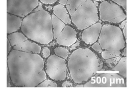
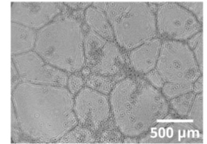
Tube length agrees to within 5%, junction counts to within 3%, and individual mesh areas correlate at Pearson r = 0.99 (n = 4).
Continuous monitoring
How the image is made
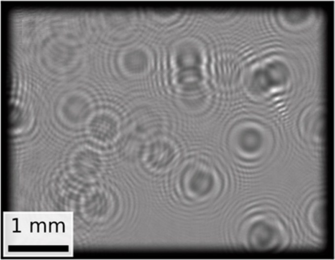
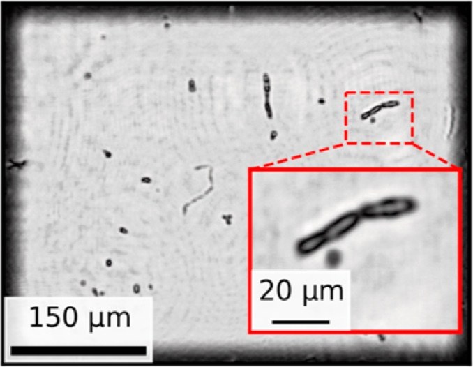
Angiogenesis panels adapted from Moncada-Madrazo et al., Biosensors and Bioelectronics 2026, 303, 118579. How the image is made, adapted from Benito-Altamirano et al., Biosensors 2025, 15(1), 40. Both are open access under CC BY 4.0. Panels were cut from the published composites and rescaled; no image content was altered.
03Published work
Peer-reviewed work by the founding team at UB, predating the company.
- [1]
Moncada-Madrazo, Canals, Caravaca-Müller, Moreno, Castro, Ramón-Azcón, Vilà, Diéguez — In-incubator continuous and endpoint monitoring of angiogenesis using a chip-sized holographic microscope — Biosensors and Bioelectronics, 303, 118579, 2026 — doi.org/10.1016/j.bios.2026.118579
- [2]
Moncada-Madrazo et al. — Chip-Sized Lensless Holographic Microscope for Real-Time On-Chip Biological Sensing — Sensors, 25(17), 5247, 2025 — doi.org/10.3390/s25175247
- [3]
Benito-Altamirano et al. — MicroVi: A Cost-Effective Microscopy Solution for Yeast Cell Detection and Count in Wine Value Chain — Biosensors, 15(1), 40, 2025 — doi.org/10.3390/bios15010040
- [4]
Diéguez et al. — Chip-Sized Microscopy for Continuous Monitoring: White Wine Fermentation and Yeast Cell Counting via Deep Learning — Eng. Proc., 78, 1, 2024 — doi.org/10.3390/engproc2024078001
- [5]
Vilà, Moreno, Canals, Diéguez — A Compact Raster Lensless Microscope Based on a Microdisplay — Sensors, 21(17), 5941, 2021 — doi.org/10.3390/s21175941
- [6]
Canals et al. — A Novel Approach for a Chip-Sized Scanning Optical Microscope — Micromachines, 12(5), 527, 2021 — doi.org/10.3390/mi12050527
- [7]
Moreno et al. — Pursuing the Diffraction Limit with Nano-LED Scanning Transmission Optical Microscopy — Sensors, 21(10), 3305, 2021 — doi.org/10.3390/s21103305
- [8]
Franch, Canals, Moro, Vilà, Prades, Diéguez et al. — Nano illumination microscopy: scanning with an array of individually addressable nanoLEDs — Optics Express, 28(13), 19044, 2020 — doi.org/10.1364/OE.391497
04Who we are
XipScope was incorporated in Barcelona in 2026 as a University of Barcelona spin-off.
Founders
Dr. Sergio Moreno
Has been developing the platform since its early stages in 2018. Combines a technical background in single-photon (SPAD) sensor design and microelectronics with company building and product strategy. Leads strategy, investor relations and operations. PhD in Engineering and Applied Sciences, UB, and MSc in Microelectronic Design.
LINKEDIN ↗
Dr. Joan Canals
Has worked in microelectronic design and optoelectronic system integration for biomedical applications since 2006. Principal architect of the platform from the outset. Leads R&D and the technical roadmap. Electronics Engineer and PhD in Engineering and Applied Sciences, UB.
LINKEDIN ↗Advisors

Mattia Nuti
More than fifteen years of experience scaling technology companies. Advises on fundraising, go-to-market and international expansion, with a focus on organ-on-a-chip in Europe and the US.
LINKEDIN ↗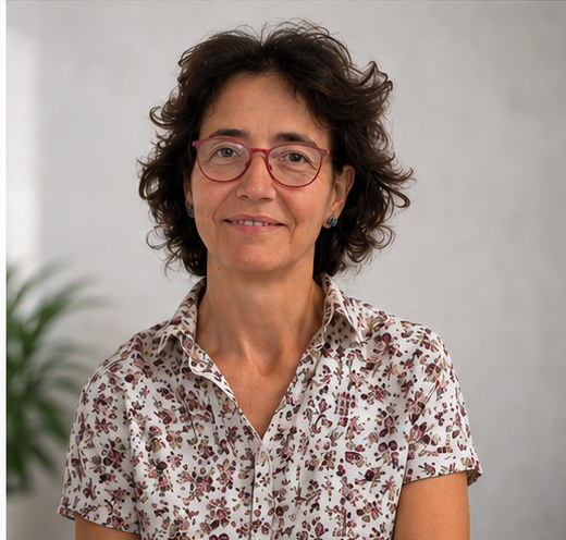
Dr. Anna Vilà
Full Professor at UB, head of the Instrumentation and Communication Systems group; computational imaging and lensless microscopy.
LINKEDIN ↗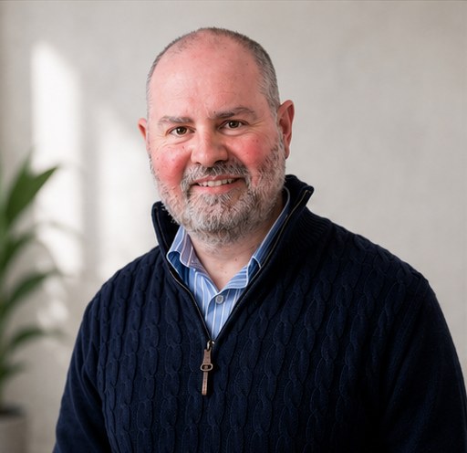
Dr. Ángel Diéguez
Full Professor at UB, head of the Microelectronic Design group; CMOS sensors and VLSI for biomedical applications.
LINKEDIN ↗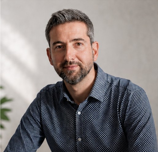
Dr. Juan Daniel Prades
Humboldt Professor at TU Braunschweig, former Full Professor at UB; semiconductor nanotechnology and microLED fabrication.
LINKEDIN ↗